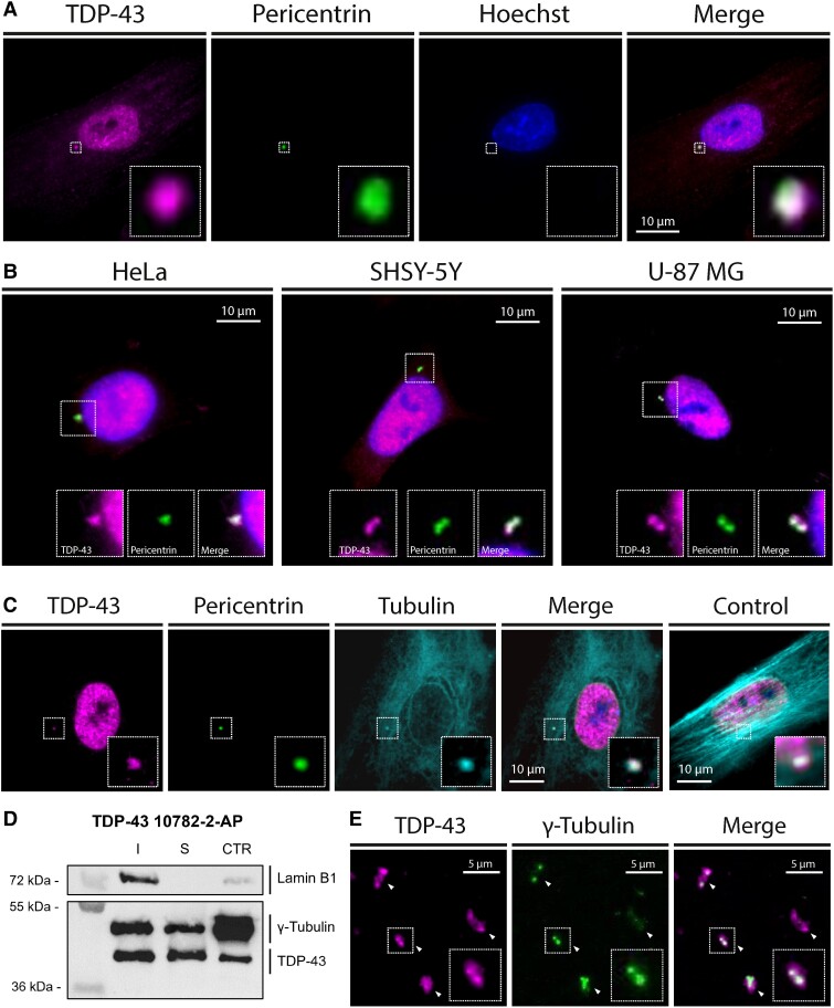
Figure 1.
Centrosomal enrichment of TDP-43 in human cells. (A and B) Total internal reflection fluorescence (TIRF) microscopy images of fixed healthy human skin fibroblasts (A), and pseudo-neuronal HeLa, U-87 MG and SH-SY5Y cells (B) immune-stained with antibodies against TDP-43 (10782-2-AP) and pericentrin, showing a co-localization of the two signals when merged. (C) Similar results were obtained in human skin fibroblasts after a 2-h treatment with nocodazole (5 mg/ml) inducing microtubule depolymerization. (D) Western blot performed on purified centrosome fractions from human T-lymphoblastoid KE-37 cells using primary antibodies against TDP-43 (10782-2-AP), γ-Tubulin and Lamin B1, showing an enrichment of TDP-43 at the centrosome. I = insoluble fraction; S = soluble fraction; CTR = centrosome fraction. (E) Immunofluorescence microscopy performed on purified centrosomes (arrowheads) fixed and immune-stained for TDP-43 (60019-2-Ig) and γ-Tubulin showing a co-localization of the two signals.

