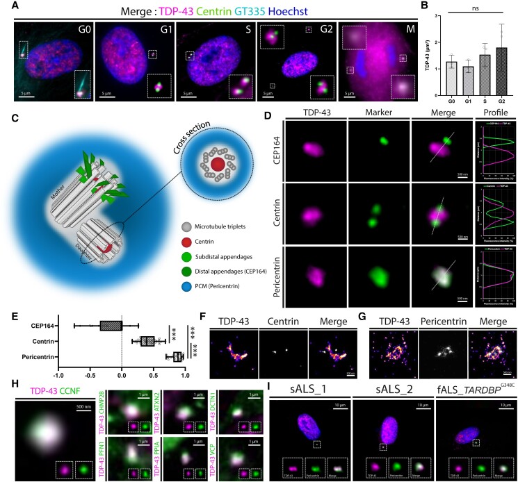
Figure 2.
Cell cycle independent pericentriolar enrichment of TDP-43 in physiological and pathological conditions. (A) Merged images of TIRF microscopy performed on human skin fibroblasts fixed and immune-stained for TDP-43, pericentrin and GT335, showing the presence of TDP-43 at the centrosome in all phases of the cell cycle, including mitosis. (B) Surface area of the TDP-43 centrosomal fraction (μm2) measured at each phase of the cell cycle (ns = not significant). (C) Schematic representation of the different parts of the centrosome, with corresponding protein markers. (D) High magnification TIRF microscopy images of the centrosome of human skin fibroblasts immune-stained with antibodies against TDP-43, centrin, CEP-164 and pericentrin. The co-localization profiles (fluorescence intensity of each channel) shown on the right and the Pearson coefficients of co-localization (E) indicate a pericentriolar enrichment of TDP-43 (P < 0.001). (F and G) Super resolution stochastic optical reconstruction microscopy (STORM) images of human skin fibroblasts immune-stained with antibodies against TDP-43 and centrin (F) or pericentrin (G). (H) TIRF microscopy images of TDP-43 and centrosomal TDP-43 partners associated to familial ALS/FTLD in fixed healthy human skin fibroblasts (magnification at the centrosome) disclosing a co-localization. (I) TIRF microscopy images of fixed human skin fibroblasts derived from sporadic and familial ALS (fALS) patients immune-stained with antibodies against TDP-43 and pericentrin, showing a co-localization of the two signals.

