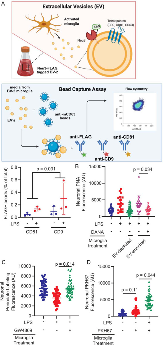
Figure 2. Neu3 is associated with microglia-derived fusogenic extracellular vesicles.

(A) Endogenous NEU3 was FLAG-tagged in BV-2 microglia by homology-directed recombination. After exposure of BV-2 microglia with endogenously FLAG-tagged Neu3 to vehicle or LPS, EVs were captured on anti-mCD81 or anti-mCD9 coupled beads, labeled with fluorophore-coupled anti-FLAG or anti-mCD63, and analyzed by flow cytometry. Bead captured-EV’s demonstrate increased FLAG signal in LPS-treated microglia compared to resting microglia, indicating that NEU3 colocalizes with EV markers and is released via EVs upon LPS-activation. (-LPS vs. +LPS, p-value = 0.031). n=3 wells/condition, 2 capture methods/well. (B) PNA staining of neuronal surfaces treated with EV-enriched or EV-depleted media of activated WT microglia reveals that EV-enriched fraction alone has sialidase activity (EV-enriched vs. EV-depleted, p=0.023; EV-enriched vs. EV-enriched+DANA, p=0.034). n=4 coverslips/condition, 7 cells/coverslip. (C) Periodate labeling of neuronal surface sialic acids reveals that pharmacologic inhibition of EV production with GW4869 abrogates sialidase activity of EV-enriched microglia media (-LPS vs. +LPS, p=0.027; -LPS vs. +LPS+GW4869, p=0.97; +LPS vs. +LPS+GW4869, p=0.014). n=3 coverslips/condition, 135 cells total. (D) Imaging of neurons treated with PKH67-stained microglial exosome demonstrates transferal of dye from EVs to neuronal membranes (Vehicle vs. -LPS, p=0.15; -LPS vs. +LPS, p=0.044). Hypothesis testing for all panels was performed using hierarchical permutation test. n=3 coverslips/condition, 15 cells/coverslip.
