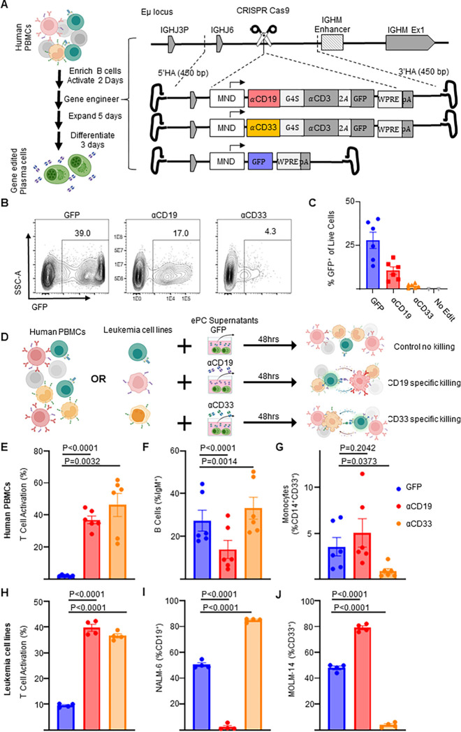
Figure 2: Human plasma cells engineered to secrete anti-leukemia bispecifics specifically target cells expressing physiological levels of antigen.
Primary human B cells were isolated and cultured for two days in activating media then edited. A) Schematic showing how primary activated human B cells were edited to express GFP or αCD19.T2A.GFP or αCD33.T2A.GFP. After editing activated B cells, the engineered cells were then cultured in expansionary media for 5 days followed by differentiation into PCs over 3 days and cells and supernatants. B) Representative flow cytometry plots assessing editing via expression of GFP and C) quantification as % of live cells. D) Schematic illustrating in vitro PBMC or Leukemia cell line killing assays. Briefly, autologous CD8+ T cells are co-cultured with PBMCs or mixed leukemia cell populations (NALM-6 and MOLM-14) in the presence of supernatants from ePCs for 48 hours. Flow cytometry was used to quantify E) T cell activation (%CD69+CD137+ of CD3+ cells), F) the % B cells (IgM+) of live cells, G) the % monocytes (CD14+CD33+) of live cells in PBMC cultures at the end of the 48-hour co-culture. Likewise flow cytometry was used to quantify H) T cell activation (%CD69+, CD137+ of CD8+ cells), the frequency of I) NAML-6 (CD19+) and J) MOLM-14 (CD33+) in the leukemia cell line killing assay. In E-G, data were obtained from six donors in three independent experiments, and in H-J, data were obtained from four donors. Error bars represent SEM. P-values were calculated using paired one-way ANOVAs with Dunnett’s posttest. Illustrations were created in part with biorender.com.

