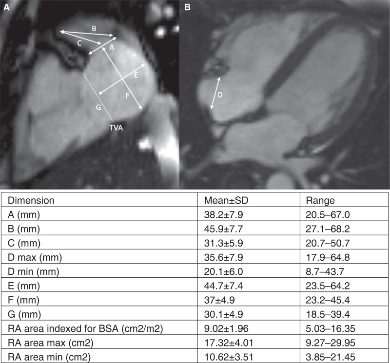
Figure 3.
Cardiac MRI dimensions analysed. Representative MRI images demonstrating the measurements taken and the resultant mean measurement; (A) two-chamber view. (B) Four-chamber view. Measurements noted: A—RAA ostium, B—RAA depth to CT, C—RAA depth to mid-point of RAA ostium, D—CT to lateral TVA, E—RA diameter (2Ch view) perpendicular to TV annulus, F—RA diameter (2Ch view) perpendicular to RAA ostium, G—TVA diameter (2Ch view). BSA, body surface area; CT, crista terminalis; MRI, magnetic resonance imaging; RA, right atrium; RAA, right atrial appendage; TVA, tricuspid valve annulus.

