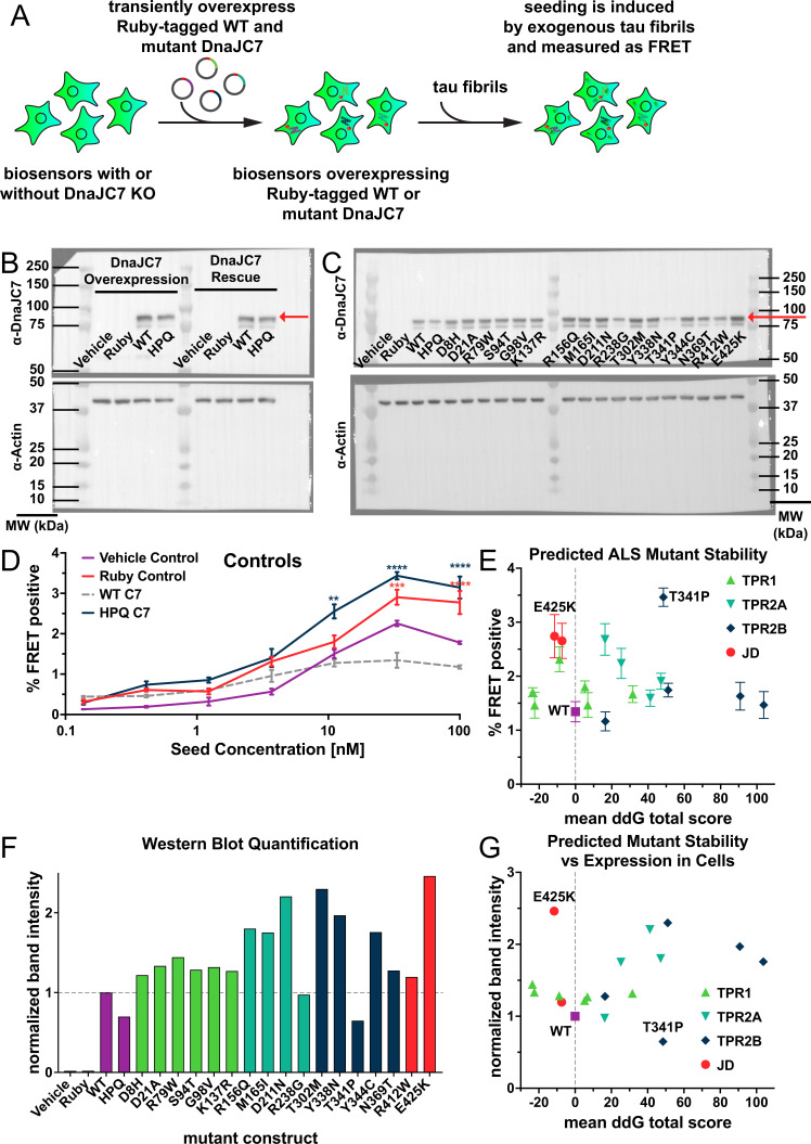
(
A) Schematic showing tau biosensor cells with or without DnaJC7 knockout (KO) transiently overexpressing different Ruby-tagged gRNA-resistant DnaJC7 constructs. Cells were allowed to express the constructs for 2 days before being plated for the seeding assay. (
B) Immunoblotting for DnaJC7 confirmed expression of the Ruby-WT DnaJC7 and Ruby-DnaJC7 (HPQ) mutant constructs in tau biosensor cells without (Overexpression) and with endogenous DnaJC7 knocked out (Rescue). Ruby fusion constructs are highlighted by a red arrow. (
C) Immunoblotting for DnaJC7 confirmed expression of the Ruby-WT DnaJC7 and Ruby-DnaJC7 amyotrophic lateral sclerosis (ALS) mutant constructs in tau biosensor cells with endogenous DnaJC7 knocked out. Ruby fusion constructs are highlighted by a red arrow. (
D) Positive and negative controls utilized in the rescue of DnaJC7 KO in tau biosensor cells with ALS-associated mutants of DnaJC7, colored as follows: Vehicle Control, purple; Ruby Control, orange; WT DnaJC7, gray dashed; HPQ mutant, dark blue. Error bars represent SEM of three technical replicates. (
E) Rosetta-calculated mean Gibbs free energy shift (ddG) of the ALS-associated mutants of DnaJC7 vs. their rescue seeding with 33nM of tau fibrils. Gray dashed line denotes a mean ddG total score of 0. Mutants are colored according to their domain localization: TPR1, green; TPR2A, teal; TPR2B, dark blue; JD, orange. (
F) Quantification of the western blot signal for the different ALS-associated mutants and controls in (
C). All constructs were normalized to the band intensity of the WT construct. Domains are colored as in (
C). (
G) Rosetta-calculated mean Gibbs free energy shift (ddG) of the ALS-associated mutants of DnaJC7 vs. their normalized western blot band intensity. Gray dashed line denotes a mean ddG total score of 0. Mutants are colored as in (
C).All error bars represent SEM of three technical seeding replicates. *=p < 0.05, **=p < 0.01, ***=p < 0.001, ****=p < 0.0001. Source data for this figure are provided in
Figure 5—figure supplement 1—source data 1.


