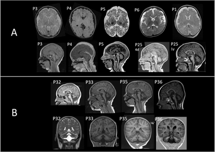
Fig. 1. Brain magnetic resonance image (MRI) of individuals with biallelic BRAT1 variants.
A Patients with RMFSL (P1, P3, P4, P5, P25). Note cerebral atrophy and enlarged subarachnoid spaces on axial and sagittal planes. B Patients with NEDCAS (P32, P33, P35, P36). Note cerebellar atrophy on sagittal and coronal planes.

