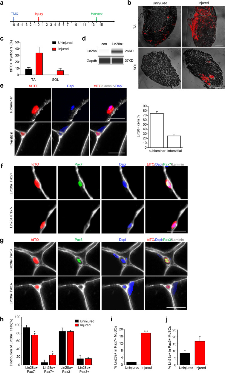
Fig. 1. Lin28a+ stem cells contribute to satellite cells and myofibers during regeneration.
a Schedule of tamoxifen (TMX) treatment. Lin28a-T2A-CreER;LSL-tdTO mice were injected with TMX, 6 days before cryoinjury, daily for the first 1 week after injury, every other day from the 7th day, and harvested on the 14th day after injury. b TA and SOL muscles of Lin28a-T2A-CreER;LSL-tdTO mice were damaged by cryoinjury and harvested on day 14 post injury. Contralateral TA or SOL muscle was used as the control. Scale bars, 600 μm. c Quantification of the number of tdTO+ muscle fibers per 100 muscle fibers in the TA and SOL muscles of Lin28a-T2A-CreER;LSL-tdTO mice on day 14 post injury and without injury. Quantification was performed on the images in b. For each muscle section, at least 3 whole sections were quantified and averaged. d Low-protein-input capillary-based immunoassays for Lin28a protein expression in Lin28a-tdTO+ and conventional (con) Pax7+ MuSCs. Gapdh protein was used as the loading control. Unprocessed original capillary-based immunoassay results are shown in Supplementary information, Fig. S7. e Cryosections of the TA muscles of Lin28a-T2A-CreER;LSL-tdTO mice that were cryoinjured and harvested on day 14 post injury. Muscle sections were co-stained for laminin (white) and DAPI (blue). Scale bars, 10 μm. Right: quantification of the percentage of sublaminar and interstitial Lin28a+ cells. For each muscle section, at least five different fields were quantified and averaged. f Cryosections of the TA muscle of Lin28a-T2A-CreER;LSL-tdTO mice at 14 days after cryoinjury. Muscle sections were co-stained for laminin (white), DAPI (blue) and Pax7 (green). Scale bar, 10 μm. g Cryosections of the TA muscle of Lin28a-T2A-CreER;LSL-tdTO mice at 14 days after cryoinjury. Muscle sections were co-stained for laminin (white), DAPI (blue) and Pax3 (green). Scale bar, 10 μm. h Quantification of the distribution of Pax3–, Pax3+, Pax7– and Pax7+ cells in the Lin28a-tdTO+ pool per field in the TA muscle of Lin28a-T2A-CreER;LSL-tdTO mice on day 14 post injury (red), relative to uninjured muscles (black). For each muscle section, at least five different fields were quantified and averaged. i Quantification of the percentage of Lin28a-tdTO+ cells in Pax7+ satellite cells per field in the TA muscle of Lin28a-T2A-CreER;LSL-tdTO mice on day 14 post injury (red), relative to uninjured muscles (black). For each muscle section, at least five different fields were quantified and averaged. j Quantification of the percentage of Lin28a-tdTO+ cells in Pax3+ MuSCs per field in the TA muscle of Lin28a-T2A-CreER;LSL-tdTO mice on day 14 post injury (red), relative to uninjured muscles (black). For each muscle section, at least five different fields were quantified and averaged. *P < 0.05, ***P < 0.001.

