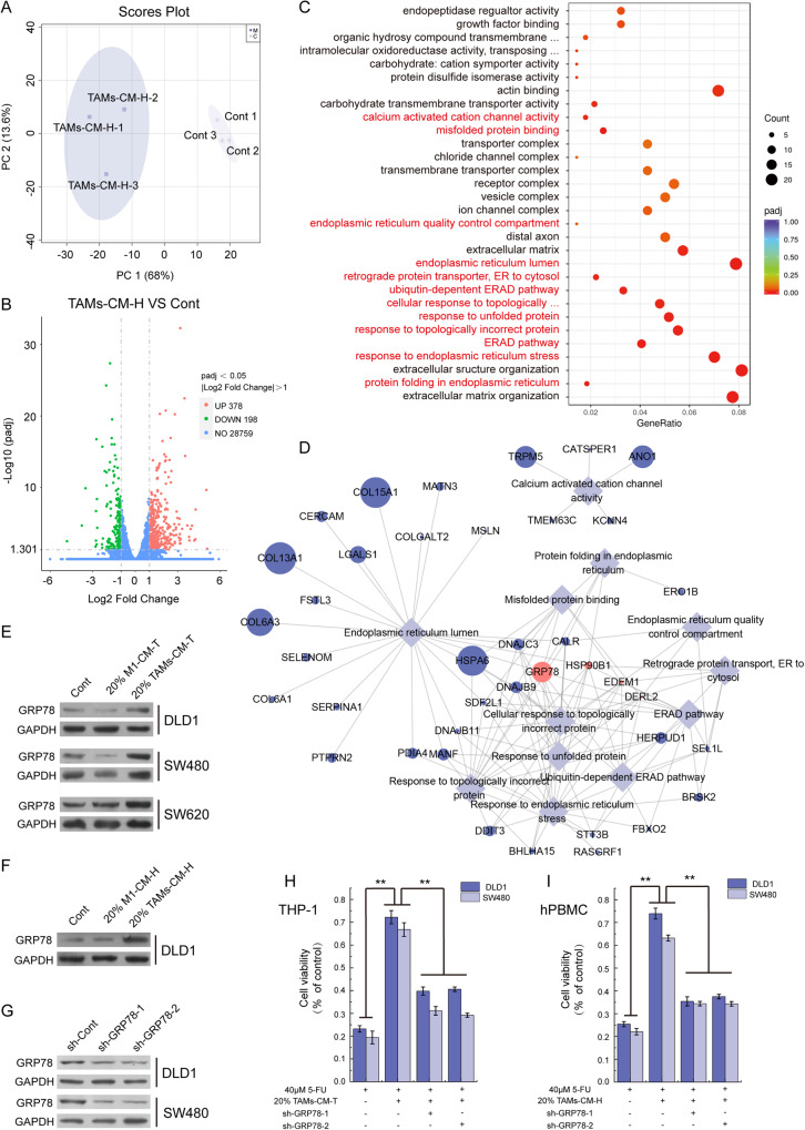
Fig. 2. GRP78 mediated the TAMs-associated CRC resistance to 5-FU.
A PCA score plot showing DLD1 (control) and TAMs-CM-H treated DLD1 (TAMs-CM-H) samples. B The volcano map illustrated the distribution of differential genes. C GO enrichment analysis of up-regulated genes (TAMs-CM-H VS Control). D The network analysis of endoplasmic reticulum-related terms and the genes involved. E After treatment with 20% M1-CM-T or 20% TAMs-CM-T, the level of GRP78 protein was detected by western blot in DLD1, SW480, and SW620 cells. F After treatment with 20% M1-CM-H or 20% TAMs-CM-H, the level of GRP78 protein was detected by western blot in DLD1 cells. G After the knockdown of GRP78 with shRNA in DLD1 and SW480 cells, the expression of GRP78 was detected by western blot. H The effect of 20% TAMs-CM-T on 5-FU resistance was detected with DLD1 and SW480 cells which with GRP78 knocking down, **p < 0.01. I The effect of 20% TAMs-CM-H on 5-FU resistance was detected with DLD1 and SW480 cells which with GRP78 knocking down, **p < 0.01.

