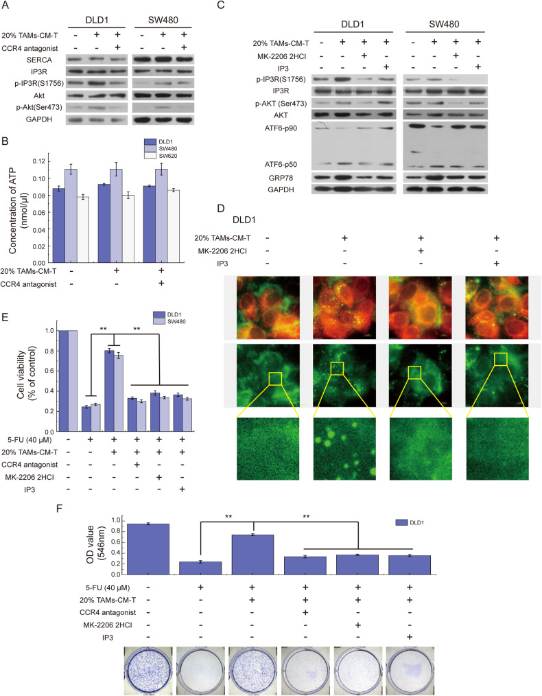
Fig. 5. IP3R inactivation induces the abnormal calcium distribution.
A SERCA, IP3R, p-IP3R, Akt, and p-Akt were detected by western blot in DLD1 and SW480 cells treated with 20% TAMs-CM-T or/and CCR4 antagonist. B The bar graph represented the intracellular total ATP concentration of DLD1, SW480, and SW620 cells treated with 20% TAMs-CM-T or/and CCR4 antagonist. C After treatment with MK-2206 2HCI, IP3, and 20% TAMs-CM-T, the expression of IP3R, p-IP3R, Akt, p-Akt, ATF6-p50, and GRP78 were detected by western blot in DLD1 and SW480. D After treatment with 20% TAMs-CM-T, MK-2206 2HCI, and IP3, DLD1 cells were fluorescence stained with Fluo-3 AM (green) and ER-tracker (red). E After blocking the CCL17/CCL22-CCR4 pathway with a CCR4 antagonist or activating IP3R with MK-2206 2HCI and IP3, the effect of TAMs-CM-T on the decreases of DLD1, SW480, and SW620 cell viability induced by 5-FU was detected by MTT, **p < 0.01. F After blocking the CCL17/CCL22-CCR4 pathway with a CCR4 antagonist or activating IP3R with MK-2206 2HCI and IP3, the effect of 20% TAMs-CM-T on the diminution of DLD1 cell colony formation capacity induced by 5-FU was detected by Colony-Formation Assay, **p < 0.01.

