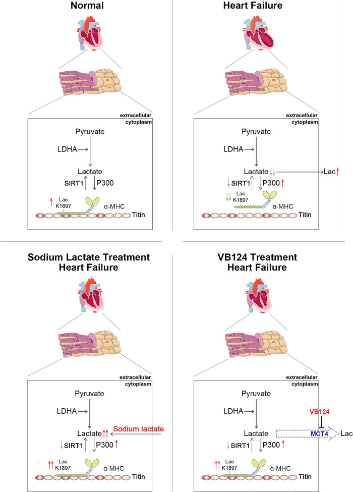
Fig. 10. Schematic diagram showing the proposed mechanistic model of α-MHC lactylation.
Under the physiological state, α-MHC lactylation reserves the interaction of α-MHC with Titin and maintains sarcomeric structure and function. Upon pathological stress stimulation, a decrease in the lactate concentration in cardiomyocytes leads to reduction of α-MHC lactylation and α-MHC–Titin interaction, thus impairing cardiac structure and function. Upregulation of the lactate concentration by administering sodium lactate or inhibiting MCT4 in cardiomyocytes can promote α-MHC lactylation and α-MHC–Titin interaction, thereby alleviating heart failure.

