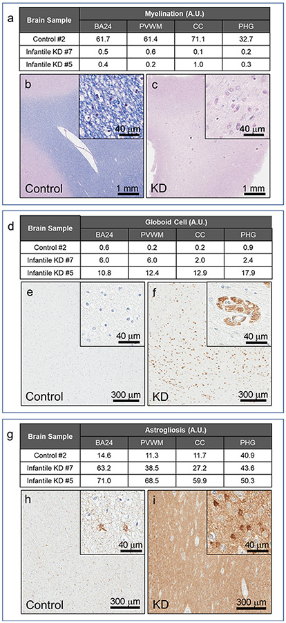
Fig. 5.
Quantitative neuropathology in the white matter regions of infantile KD. Neuropathological features of infantile KD are illustrated in two cases (cases #5 and #7) compared to an age-matched normal control (case #2). White matter regions (BA24, PVWM, CC and PHG) are analyzed for levels of myelination, globoid cells and astrogliosis with Luxol fast blue stains (a-c), CD68 immunohistochemistry (d-f) and GFAP immunohistochemistry (g-i), respectively. Representative images of LFB-PAS stain (b, c), CD68 (e, f) and GFAP (h, i) in BA24 white matter region of control and KD cases are shown with results of quantification (a, d, g).

