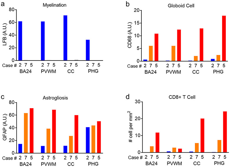
Fig. 7.
Disease severity-dependent increase of neuropathology in the white matter regions of infantile KD brains. Image quantification results for myelination, globoid cells, astrogliosis and CD8+ T cells in white matter (BA24, PVWM, CC, PHG) of a control (case #2, blue) and of two infantile KD cases with different disease severity (case #7, orange; more severe case #5, red) are plotted in graphical representations.

