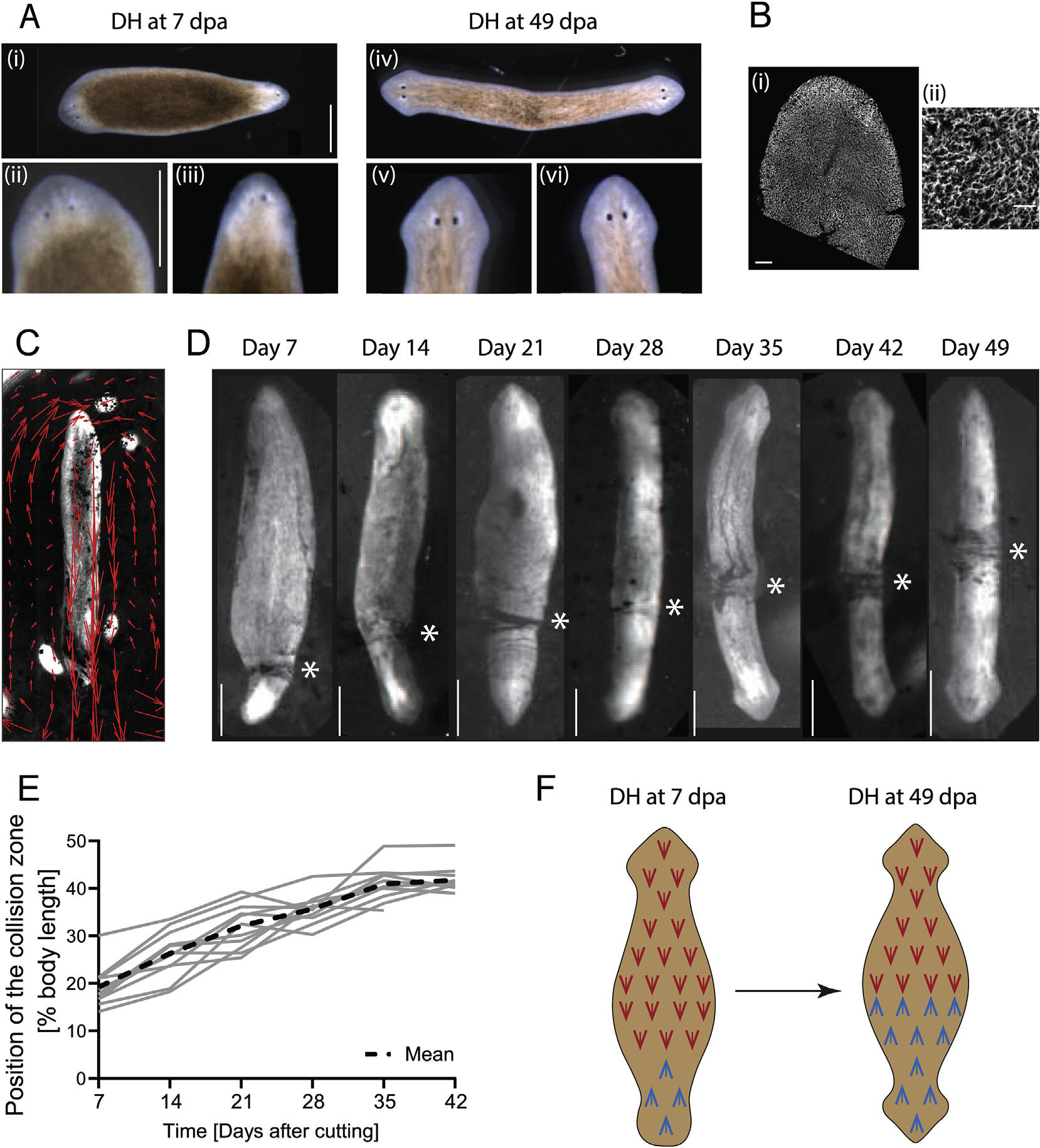
Fig. 1. Flow driven by the multiciliated epithelium can be tracked and epithelial polarity dynamically remodels in DH animals.

A) (i) Morphological differences in head development in DHs can be observed at Day 7 (days after amputation – dpa), revealing that the primary head (ii) is more developed than the secondary head (iii). (iv) In DHs at Day 49 both heads (v, vi) are equally developed. Scale bars 1 mm. B) Cilia of the multiciliated ventral surface epithelium stained with antibody against acetylated-tubulin at (i) low and (ii) high magnification. Scale bar 100 μm in (i) and 25 μm in (ii). C) Flow driven by cilia beat visualized with carmine powder and tracked with particle image velocimetry (PIV) in a single-headed animal (red arrows). D) Opposing flow directions in DH animals lead to the accumulation of particles at the point where the opposing flow fields meet. This “collision zone” (asterisk) repositions towards the middle of the animal from Day 7 until Day 49. E) Plot of position of the collision zone as percentage of total body length for 12 animals individually tracked over time, measured every 7th day, and mean curve (dashed line). N = 12, with 5 repeats showing similar pattern. F) Schematic representing the change in cilia orientation in the posterior half of the DH as the cilia orientation changes over time.
