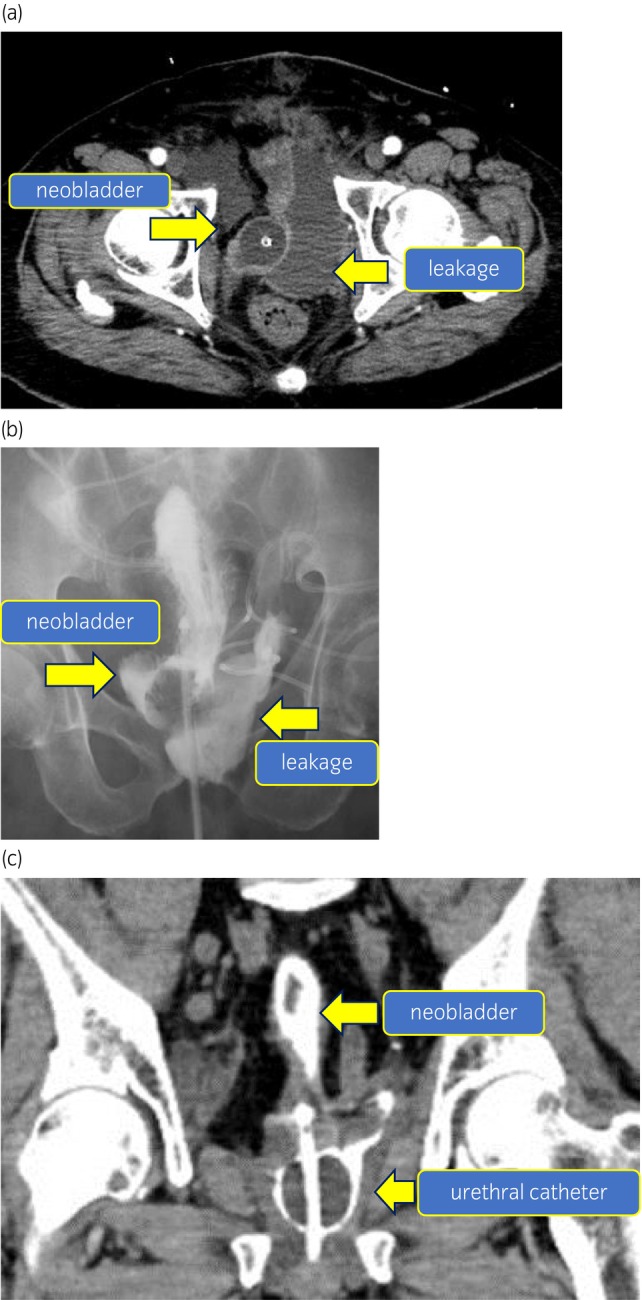
Fig. 1.

(a) CT image showing the leakage. CT scan of horizontal view for the fever examination revealed large amount of fluid collection on the left side of the urethral‐neobladder anastomosis. This may be a lymphocele infection or leakage of urine. (b) Image of leakage in cystography. Cystography reveals a large amount of leakage on the left side of the anastomosis. It reveals a neobladder‐urothelial anastomotic leakage. (c) CT image of severe leakage. CT scan of the coronary view showing urethral catheter falling off. The urethral balloon is falling off from the neobladder in a CT scan of coronary view. It suggests a severe anastomotic dehiscence of suture.
