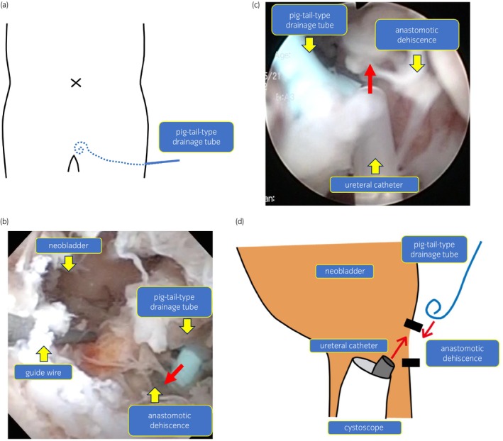
Fig. 2.

(a) Image of drainage tube position. We placed a pigtail‐type drainage tube transperineally beside the anastomosis of the neobladder. (b) Image of injecting fibrin glue with a cystoscope. We injected 10 mL of fibrin glue into the large space beside the anastomotic dehiscence from the drainage tube. (c) Image of injecting fibrin glue with a cystoscope. We sprayed 5 mL of fibrin glue around the anastomotic dehiscence from the ureteral catheter using a cystoscope. (d) Schematic diagram of injecting fibrin glue. First, we injected fibrin from the drainage tube. Second, we injected fibrin glue from the ureteral catheter using a cystoscope.
