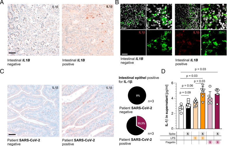
FIGURE 3.
IL-1β can be produced by epithelial cells and macrophages in the intestine during SARS-CoV-2 infection. (A) Representative picture of IL-1β expression, determined by immunohistochemistry, in the duodenum of patients with nondetectable intestinal IL1B levels (left) and detectable intestinal IL1B levels (right). Tissue samples were taken during the autopsy. Counterstaining and bluing were performed with hematoxylin and Bluing Reagent. Scale bar, 50 µm. (B) Representative picture of IL-1β (red) and IBA1 (green) expression, determined using double fluorescence, in the duodenum of patients with nondetectable intestinal IL1B levels (left four pictures) and detectable intestinal IL1B levels (right four pictures). The upper panel represents merged pictures; the lower panel represents unmerged channels. Tissue samples were taken during autopsies. Scale bar, 50 µm; closeup, 20 µm. (C) Left: Representative picture of IL-1β expression, determined by immunohistochemistry, in the duodenum of patients without SARS-CoV-2 diagnosis (far left, n = 3) and with clinical SARS-CoV-2 diagnosis (middle, n = 3). Tissue samples were taken during surgery. Counterstaining and bluing were performed with hematoxylin and Bluing Reagent. Scale bar, 50 µm. Right: Percentage of patients with detectable IL-1β expression in the intestinal epithelium. (D) IL-1β expression of human intestinal organoids (n = 6), measured in supernatants by ELISA. Suspension organoids were prepared and placed in a differentiation medium for 6 d. Organoids were then stimulated with SARS-CoV-2 S-protein (Spike) (1 µg/ml), LPS (100 ng/ml), and/or flagellin (100 ng/ml) for 10 h. Data are represented as six biological replicates. Horizontal lines represent the mean ± SEM; each symbol indicates one biological replicate from a different patient.

