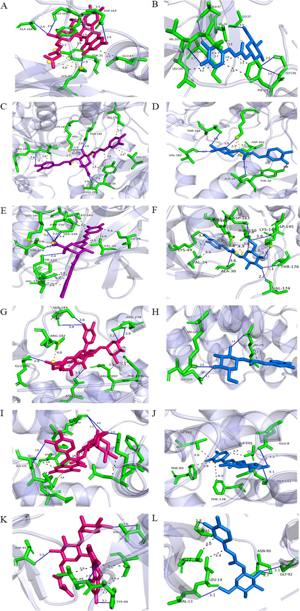
Fig. 8.
The in silico interactions between selected signalling protein molecules and polyphenol ligands. A and B p38α complex, (C and D) ERK complex, (E and F) JNK complex, (G and H) NF-κB complex, (I and J) Calmodulin-1 complex, (K and L) PKCα complex. Amino acid residues involved in interaction were shown in green, quercetin glucoside ligand was shown as pink structure, 3,5-di-O-caffeoylquinic acid was shown as purple structure and caffeoyl-D-glucose ligand was shown as blue structure. Hydrogen bonds were represented by blue lines, and hydrophobic interactions were represented by grey dotted lines

