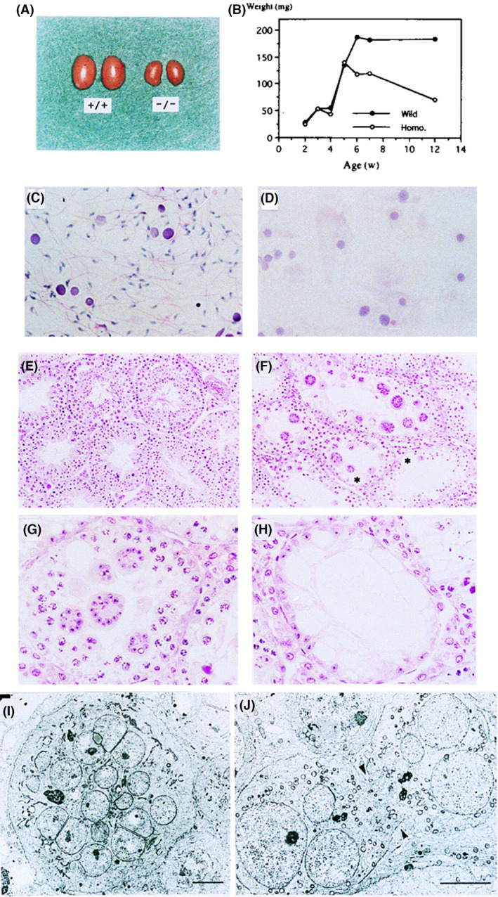
Fig. 2.

Aspermatogenesis and male sterility in GM2/GD2 synthase KO mice. Morphology and growth of the testis of WT mice and the KO mice. (A) Eight‐week‐old WT (+/+) and the KO (−/−) testes. A bar indicates 5 mm. (B) Changes in testicular weight in WT and KO mice. (C, D) Smear of seminiferous fluid from WT (C) and KO (D) mice. (Hematoxylin/eosin) (E, F) Histopathology of testis from 10‐week‐old WT (E) and KO (F) mice. (H/e) (G, H) High magnification of KO mice testis (H/e) Note the diffuse vacuoles in Sertoli cells. Bars in C, D, G, and H indicate 25 μm, and bars in E and F indicate 50 μm. (I, J) Electron micrographs of multinuclear giant cells. A giant cell (I) and an unseparated prematurely opened intercellular bridge (arrows) (J) are shown. Bars indicate 5 μm. This figure is reproduced from our previous paper [26].
