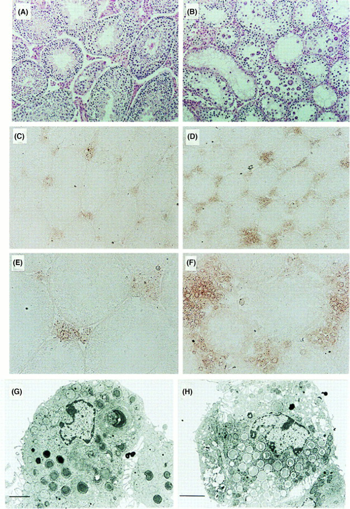
Fig. 4.

Accumulation of testosterone in the interstitial cells of GM2/GD2 synthase KO mice. Testosterone production in the interstitial cells of testis. (A, B) Hematoxylin/eosin staining of testis from wild‐type (A) and the KO (B) mice. (C–F) Immunohistochemistry for testosterone with polyclonal antibody in wild‐type (C, E) and the KO (D, F) testis. Bars in A–D indicate 50 μm, Bars in E and F indicate 25 μm. (G, H) Electron micrograph of Leydig cells of wild‐type (G) and the KO (H) mice. Bars indicate 2 μm. This figure is reproduced from our previous paper [26].
