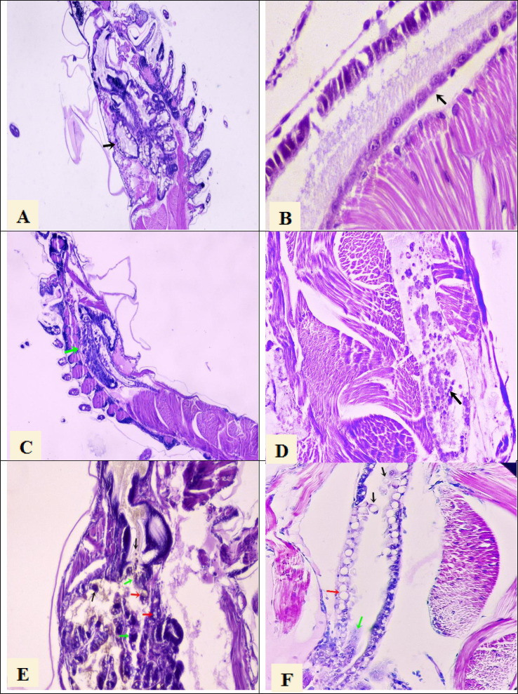
Figure 1. Histological section of P. vannamei shrimp postlarvae stained with Mayer-Bennet haematoxylin and eosin.
(A) The structure of the tubules of the hepatopancreas is observed under normal conditions, black arrow, 4x. (B) The midgut epithelium is observed in normal conditions, black arrow, 10x. (C) Necrosis of the hepatopancreatic tubules show a terminal phase of AHPND with the destruction of epithelial cells, green arrow. 4x. (D) Necrosis and detachment of epithelial cells of the hindgut into its lumen, black arrow. 10x. (E) Hepatopancreatic tubules show cell detachment, black arrows, necrosis of the tubules, green arrows, and vacuolization in the tubules caused by bacterial infection, red arrow. 10x. (F) Detachment of hindgut epithelial cells into the lumen, black arrows, necrosis in the epithelium, green arrow, and vacuolization due to bacterial infection, red arrow. 10x.

