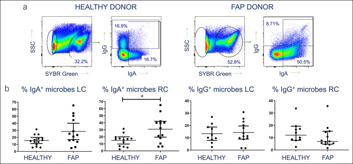
Figure 3.
Increased IgA response to intraepithelial colonic microbes in FAP. (a) Representative flow cytometry profiles showing identification of IEM by DNA staining, followed by analysis of IgA and IgG antibody coating of bacteria, in a healthy control (left) and an FAP donor (right). (b) Pooled data from all donor left and right colon samples, showing proportions of IEM coated with IgA and IgG. Median values ± 95% confidence intervals are shown; statistically significant differences between groups (Mann-Whitney tests) are indicated. FAP, familial adenomatous polyposis; IEL, intraepithelial lymphocytes; IEM, intraepithelial microbe.

