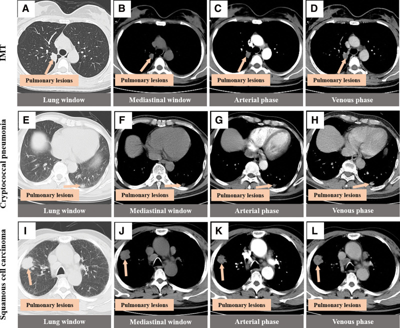
Figure 4.
CT findings of three different lung masses note. (A–D) Oval soft tissue density nodules in the right upper lung, with shallow lobulated edges, mild to-moderate uneven delayed enhancement on enhanced scanning, and slight adjacent pleural thickening. (E–H) Oval soft tissue density nodules in the left lower lung with blurred boundary and obvious uniform enhancement on enhanced scan. (I–L) Circular soft tissue density nodules in the right upper lung with deep lobulated edges, marked uneven enhancement on contrast-enhanced scan with small areas without enhancement. CT = computed tomography.

