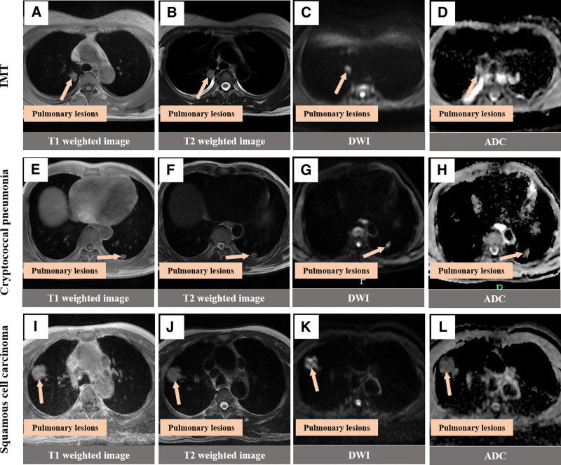
Figure 5.
MRI findings of three different types of lung masses Note. (A–D) The T1 and T2 signals of oval shape in the right upper lung were slightly longer, with high signal on DWI and high signal on ADC. (E–H) In the left lower lung, the T1 signal was slightly longer than the T2 signal, the DWI showed a high signal, and the ADC showed a slightly high signal. (I–L) The right upper lung is round, equal to T1 and slightly longer T2 signal, high signal on DWI and low signal on ADC. ADC = apparent diffusion coefficient, DWI = diffusion-weighted imaging, MRI = magnetic resonance imaging.

