. 2023 Aug 22;308(2):e230344. doi: 10.1148/radiol.230344

Figure 10:

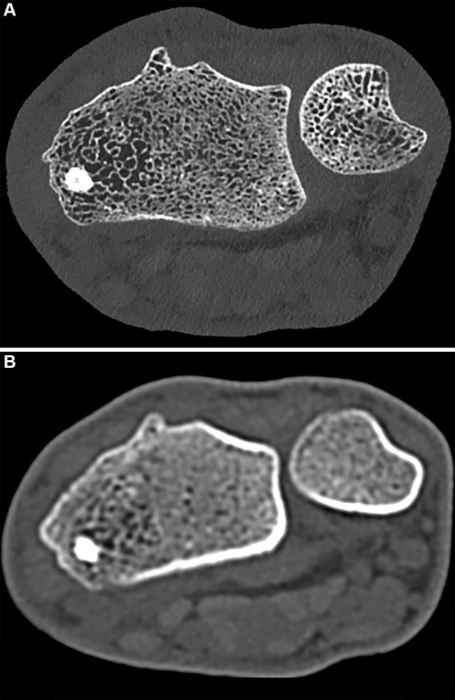
Noncontrast CT images of the distal radius in a 23-year-old male patient. (A) Ultrahigh-resolution axial photon-counting CT image (section thickness, 0.2 mm; reconstruction kernel, Br84; 120 kV; volume CT dose index, 12 mGy; window width, 500 HU; window level, 2000 HU) and (B) standard-resolution CT scan in the same patient acquired with an energy-integrating detector CT system 5 years earlier (section thickness, 1 mm; reconstruction kernel, B70; 130 kV; volume CT dose index, 15 mGy; window width, 500 HU; window level, 2000 HU; Siemens Emotion 16) show significant difference in visual resolution.

Noncontrast CT images of the distal radius in a 23-year-old male patient. (A) Ultrahigh-resolution axial photon-counting CT image (section thickness, 0.2 mm; reconstruction kernel, Br84; 120 kV; volume CT dose index, 12 mGy; window width, 500 HU; window level, 2000 HU) and (B) standard-resolution CT scan in the same patient acquired with an energy-integrating detector CT system 5 years earlier (section thickness, 1 mm; reconstruction kernel, B70; 130 kV; volume CT dose index, 15 mGy; window width, 500 HU; window level, 2000 HU; Siemens Emotion 16) show significant difference in visual resolution.