. 2023 Aug 22;308(2):e230344. doi: 10.1148/radiol.230344

Figure 2:

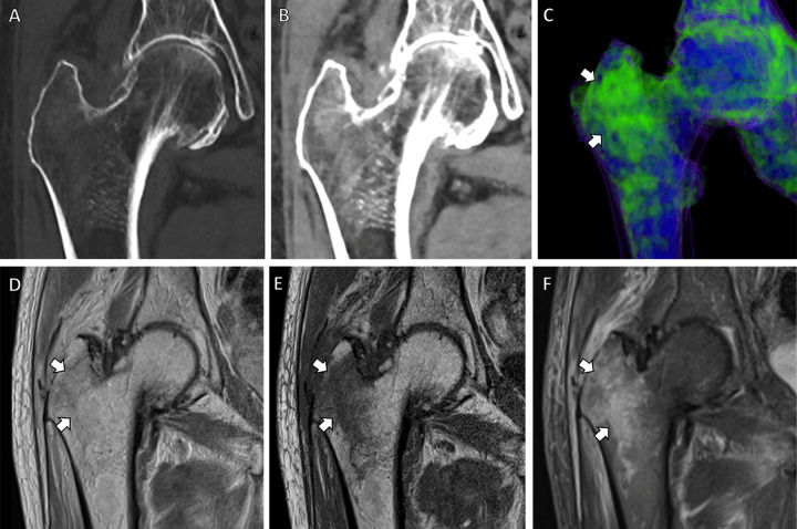
Dual-energy CT–based virtual noncontrast coronal images in an 83-year-old male patient with right-sided hip pain. (A, B) CT scans obtained with bone (A) and soft-tissue (B) windows show bone marrow edema in the right greater trochanter. (C) Bone marrow edema map redemonstrates these findings in precise detail (arrows). Corresponding (D) coronal proton density, (E) coronal T1-weighted, and (F) coronal T2-weighted fat-suppressed MRI scans demonstrate a nondisplaced microtrabecular fracture (arrows) in the same region.

Dual-energy CT–based virtual noncontrast coronal images in an 83-year-old male patient with right-sided hip pain. (A, B) CT scans obtained with bone (A) and soft-tissue (B) windows show bone marrow edema in the right greater trochanter. (C) Bone marrow edema map redemonstrates these findings in precise detail (arrows). Corresponding (D) coronal proton density, (E) coronal T1-weighted, and (F) coronal T2-weighted fat-suppressed MRI scans demonstrate a nondisplaced microtrabecular fracture (arrows) in the same region.