. 2023 Aug 22;308(2):e230344. doi: 10.1148/radiol.230344

Figure 5:

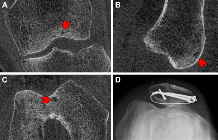
Images in a 68-year-old male patient 9 months after open reduction and internal fixation for a patellar fracture. Cone-beam CT scans in the (A) coronal, (B) sagittal, and (C) axial planes show a small subchondral cyst (arrowhead). (D) Skyline view radiograph of the same knee demonstrates the hardware used for open reduction and internal fixation; however, no subchondral cyst is detectable on the radiograph.

Images in a 68-year-old male patient 9 months after open reduction and internal fixation for a patellar fracture. Cone-beam CT scans in the (A) coronal, (B) sagittal, and (C) axial planes show a small subchondral cyst (arrowhead). (D) Skyline view radiograph of the same knee demonstrates the hardware used for open reduction and internal fixation; however, no subchondral cyst is detectable on the radiograph.