. 2023 Aug 22;308(2):e230344. doi: 10.1148/radiol.230344

Figure 8:

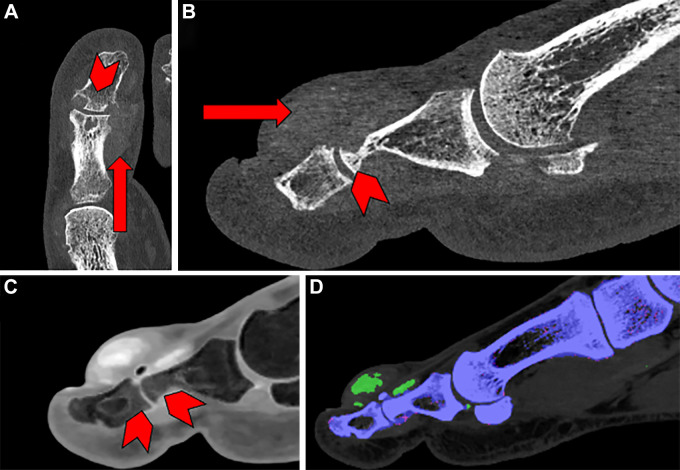
Images in a 58-year-old male patient with pain and swelling of the first digit of the left foot acquired using photon-counting detectors (PCDs). (A) Transverse view and (B) sagittal view show that periarticular mineralization (arrow) can be seen with associated erosive change (arrowhead). (C) Material decomposition acquisition reveals bone edema in the phalanges of the interphalangeal joint (arrowheads). (D) Another color-encoded material decomposition image reveals monosodium urate crystal deposits (green).

Images in a 58-year-old male patient with pain and swelling of the first digit of the left foot acquired using photon-counting detectors (PCDs). (A) Transverse view and (B) sagittal view show that periarticular mineralization (arrow) can be seen with associated erosive change (arrowhead). (C) Material decomposition acquisition reveals bone edema in the phalanges of the interphalangeal joint (arrowheads). (D) Another color-encoded material decomposition image reveals monosodium urate crystal deposits (green).