Figure 6.
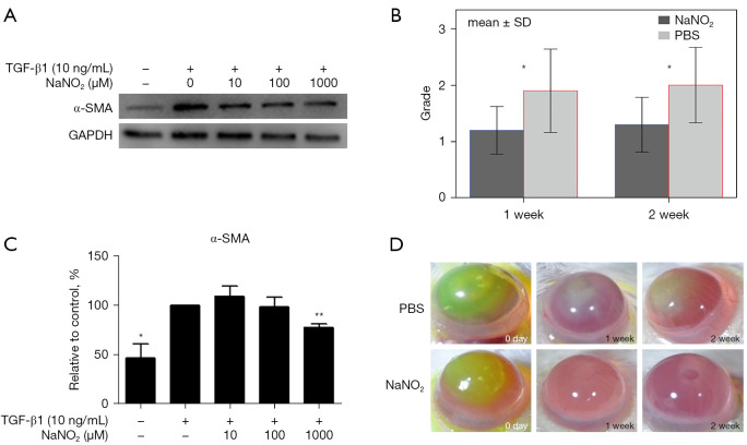
NO effect on corneal wound healing. NaNO2 was used as an NO donor. (A) Cultured human keratocytes were stimulated by transforming growth factor β1 and myofibroblast differentiation was verified by increased α-SMA expression in protein blots. Addition of NaNO2 in the culture media inhibited myofibroblast differentiation of keratocytes, as verified by decreased α-SMA expression. Approximately 20% decreased α-SMA expression was observed when 1,000 µM of NaNO2 was added to the medium (unpublished data by the authors). (B) In vivo effect of NO in corneal chemical burn in Balb/c mice. Corneal opacity grade after healing from chemical burns significantly decreased with the topical treatment of NaNO2 compared with PBS treatment. (C) The expression of α-SMA in keratocytes after TGF-β1 stimulation is attenuated by topical NaNO2 treatment. (D) Representative ocular surface pictures of healing process of chemical burn shows more transparent cornea in NaNO2-treated mouse compared with PBS control. Panels B and C are from Fig. 6 in “Effect of Nitric Oxide on Human Corneal Epithelial Cell Viability and Corneal Wound Healing” Park et al. Sci Rep. 2017;7(1):8093 under Creative Commons Attribution 4.0 International License. *, P<0.05; **, P<0.01. TGF-β1, transforming growth factor beta-1; GAPDH, glyceraldehyde-3-phosphate dehydrogenase; SD, standard deviation; PBS, phosphate buffered saline; α-SMA, alpha smooth muscle actin; NO, nitric oxide.

