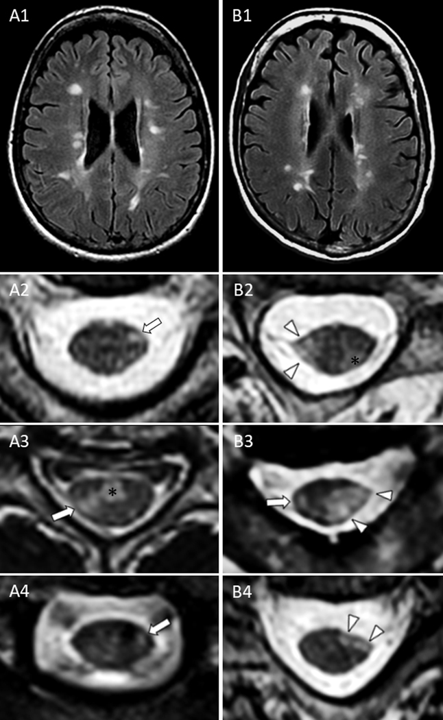
Figure 2 – Representative MRI examples of brain lesions and spinal cord lateral column lesions in patients with RRMS (left column) and SPMS (right column).

Despite a similar brain lesion burden on axial fluid attenuated inversion recovery (A1-B1), cervical spine lateral column lesions in patients with RRMS were typically characterized by smaller foci of T2-hyperintensity (less likely to involve the lateral corticospinal tracts) without atrophy on axial images (A2-A4, arrows) that differed from the large, markedly atrophic lesions of SPMS patients (B2-B4, arrowheads). The asterisks (*) indicate additional ventral (A3) and dorso-lateral (B2) prominent demyelinating lesions similarly noted in both groups, but located outside the corticospinal tracts and therefore unlikely to contribute to motor progression.
