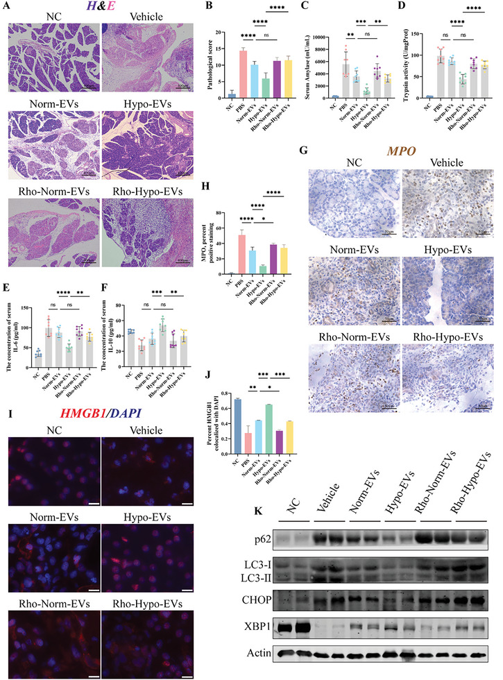
Figure 2.

Mitochondria play a key role in the treatment of SAP with hypo‐MSC‐EVs. A) H&E stained of pancreas slices in different treatment groups, scale bar: 400 µm. B) Pathological scores for H&E stained in the pancreas. Concentration of C) amylase, E) IL‐6, and F) IL‐10 in the serum. D) The level of trypsin activity in the pancreas. G,H) Immunostained of pancreatic tissue for the neutrophil marker myeloperoxidase (MPO) to measure the degree of inflammatory infiltration in the scored pancreas. I,J) Subcellular localization of immunofluorescently labelled HMGB1 in pancreatic alveolar cells and statistical analysis using “coloc2,” scale bar: 50 µm. K) Markers/mediators in endoplasmic reticulum (ER) stress and autophagy pathways in pancreatic tissue were analyzed by WB. *p < 0.05, **p < 0.01, ***p < 0.001, and ****p < 0.0001.
