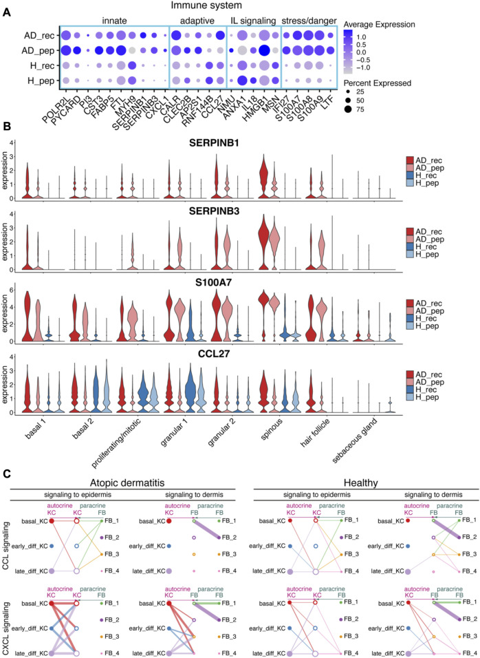
FIGURE 3.
House dust mite allergen Der p 2 initiates inflammatory pathways in KC. (A) Bubble plot depicting the expression of immune system-relevant genes in KC from skin exposed to Der p 2 protein (AD_rec, H_rec) and Der p 2 peptides (AD_pep, H_pep). Blue rectangles highlight genes relevant for the innate IS, the adaptive IS, interleukin (IL), and stress/danger signaling. (B) Violin plots show the average gene expression in KC clusters from AD_rec (dark pink), AD_pep (light pink), H_rec (dark blue), and H_pep (light blue) skin samples (n = 4). (C) Analysis of signaling crosstalk via soluble and membrane-bound factors in KC and FB. Hierarchy plots with the signal source plotted left for autocrine signaling (pink) and right for paracrine signaling (green) are shown. The receiving cell subsets (signal target) were plotted in the middle (left plot: signaling to epidermis; right plot: signaling to dermis). The plots illustrate the probability of cell–cell communications in AD and H for CCL (top panel) and CXCL signaling pathways (lower panel). Thick lines represent high probability of cell–cell interactions. AD, atopic dermatitis; H, healthy; FB, fibroblast; IL, interleukin; IS, immune system; KC, keratinocyte; rec, recombinant; pep, peptide.

