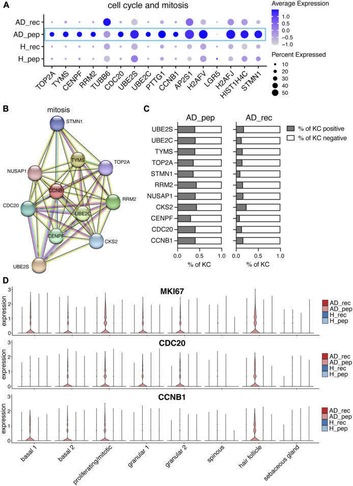
FIGURE 5.
Der p 2 peptides upregulate KC hyperproliferation in AD. (A) Bubble plot showing the expression of cell cycle and mitotic genes in KC from skin exposed to Der p 2 protein (AD_rec and H_rec) and Der p 2 peptides (AD_pep and H_pep). (B) Functional enrichment analysis identified mitotic genes enriched in AD skin exposed to Der p 2 peptides. Genes identified by differential gene expression between AD_pep and AD_rec were further analyzed using the STRING network database. Identified genes are visualized by circles and their predicted associations with lines. (C) Percentage of KC-expressing genes identified with STRING is shown in (B) for AD skin exposed to Der p 2 peptides (pep) and recombinant protein (rec). (D) Violin plots show the average gene expression in KC clusters from AD_rec (dark pink), AD_pep (light pink), H_rec (dark blue), and H_pep (light blue) skin samples (n = 4 per group). AD, atopic dermatitis; H, healthy; KC, keratinocyte; rec, recombinant; pep, peptide.

