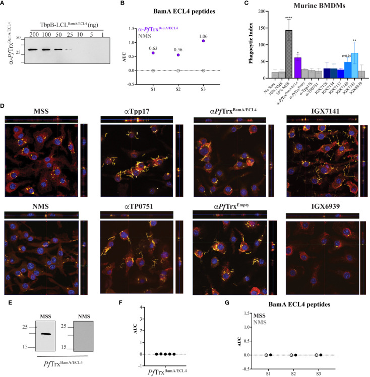
Figure 5.
Identification of an opsonic BamA ECL4 mAb. (A) Immunoblot reactivities of pooled sera (diluted 1:1,000) from five mice hyperimmunized with PfTrxBamA/ECL4 against graded nanogram amounts of TbpB-LCLBamA/ECL4. (B) ELISA reactivity of murine PfTrxBamA/ECL4 antisera or NMS with native S1, S2, and S3 peptides represented as AUC values. (C) Freshly extracted Tp were pre-incubated with 10% heat-inactivated NMS, pooled MSS, mouse antisera to PfTrxBamA/ECL4, PfTrxEmpty, TP0751 or Tpp17, or 10 μg/ml of the individual mAbs followed by incubation with murine BMDMs for 4 h at an MOI 10:1. Phagocytic indices were determined as described in Materials and methods. Asterisks show significant differences with p-values of ≤0.05, ≤0.01, or <0.0001. (D) Each representative confocal micrograph is a composite of 9–12 consecutive Z-stack planes with labeling of Tp, plasma membranes, and nuclei shown in green, red, and blue, respectively. (E) Immunoblot reactivity of pooled MSS and NMS (diluted 1:250) against 200 ng of PfTrxBamA/ECL4. ELISA reactivity (AUC values) of pooled MSS against (F) PfTrxBamA/ECL4 and (G) the S1, S2, and S3 peptides.

