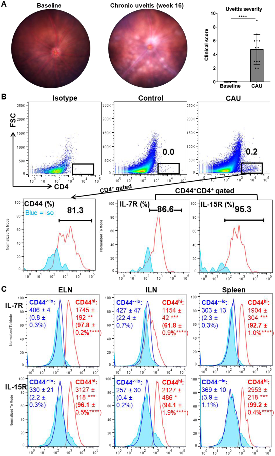
Figure 1. Chronic uveoretinitis is characterized by memory-phenotype CD4+ T cells infiltrated in the retina and present in secondary lymphoid organs (SLO).

(A) Representative digital fundus images from one same animal before disease induction (baseline) and 16 weeks post-induction (CAU). Chronic uveitis is characterized by optic disc inflammation with masked vessels, blood vessels cuffing, and multiple retinal solitary, small lesions, and disease severity is scored and summarized in the bar graph. (B) Pooled retinal tissues of each group (n = 4) from one representative experiment were analyzed by flow cytometry. CAU exhibits considerable CD4+ T cell infiltration in retinal tissues, and they are phenotyped as dominant CD44+/hi cells which are also primarily L-7R+IL-15R+ cells. Numbers in the upper panel indicate percentages among total retinal cells and in the lower panel indicate percentages among parent CD4+ (for CD44) or CD44+CD4+ (for IL-7R and IL-15R) populations. (C) The cervical eye-draining lymph nodes (ELN), the inguinal lymph nodes (ILN), and spleen in CAU mice were analyzed for IL-7R and IL-15R expressions by CD44hiCD4+ memory T cells and CD44−~loCD4+ cells. Numbers in histograms indicate mean ± SEM of MFI and percentage (in the brackets) of indicated molecules (n = 4 per group). Data shown are one representative out of two performed. *, p < 0.05; **, p < 0.01; ***, p < 0.001; ****, p < 0.0001.
