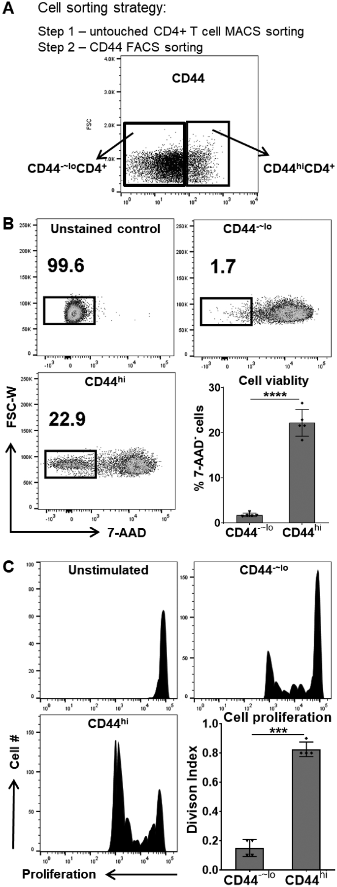
Figure 2. Chronic uveitis-derived memory T cells exhibit better survival and increased proliferative capacity.

(A) The CD44hiCD4+ memory T cells and the CD44−~loCD4+ control T cells were sorted from mice with CAU. (B) An equal number of live, sorted cells were in vitro cultured for 5 days and stained with 7-AAD, and the viable cells were determined as 7-AAD unstained populations by flow cytometric analysis. (C) The sorted cells were also in vitro stimulated with the uveitogenic antigen (IRBP) for 3 days. Cell proliferation was detected by CFSE dilution, and T cell Division Index (the average number of cell divisions that a cell in the original population has undergone) was determined by flow cytometric analysis. No antigen-stimulated CD44hiCD4+ cell cultures served as the unstimulated control. Data in bar graphs represent mean ± SEM from one experiment out of two performed. ***, p < 0.001; ****, p < 0.0001.
