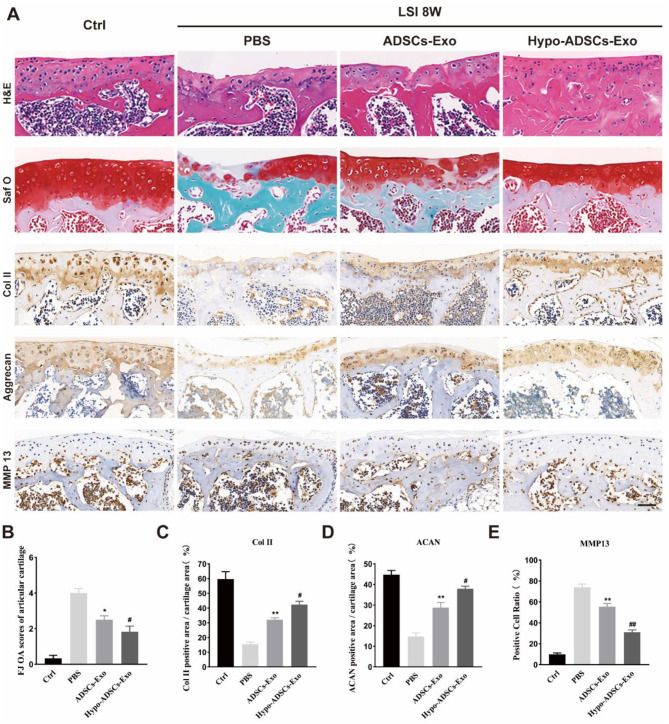
Fig. 4.
Hypo-ADSCs-Exo protect lumbar facet joint cartilage from degradation. (A) Representative histological images of LFJ cartilage with Hematoxylin-eosin (H&E) and Safranin O/Fast Green (top two) at 8 weeks post operation. Representative immunohistochemistry images of Collagen II, Aggrecan (middle two), and matrix metallopeptidase 13 (MMP13) (bottom) of LFJ cartilage. All images were captured under 40x objective lens. Scale bar=50 μm; (B) Semi-quantitative analysis of FJ OA scores of articular cartilages in (A); (C-E) Quantitative analysis of col II, Aggrecan, MMP-13 LFJ articular cartilage at 8 weeks post operation. All data are shown as the mean ±standard deviation (SD). n=6 per group. **p < 0.01, compared with PBS treated group mice, #p < 0.05, ##p < 0.01 compared with ADSCs-Exo mice. n=6 per group

