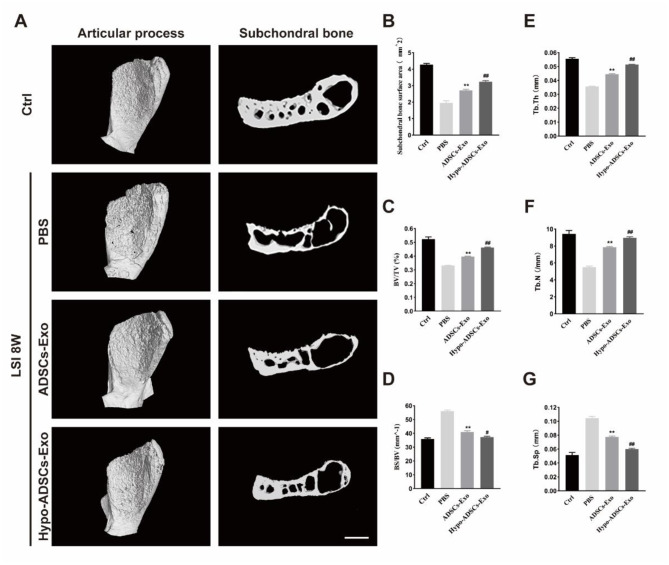
Fig. 6.
Hypo-ADSCs-Exo preserved the subchondral bone microarchitecture in LFJ OA. (A) 3D reconstructed X-ray microscope images of sub-chondral bone surface of the superior articular process and (left panel) corresponding sagittal microstructure (right panel) among the control, PBS or ADSCs-Exo and Hypo-ADSCs-Exo treated groups. Scale bar=200 μm; (B) – (G) Histomorphometry analysis of 3D images of the LFJ subchondral bone among four different groups. All data are shown as the mean ±standard deviation (SD). n=6 per group. **p<0.01, compared with PBS treated group mice, #p < 0.05, ##p < 0.01, compared with ADSCs-Exo mice

