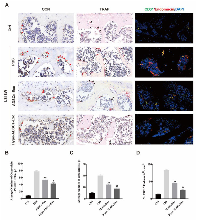
Fig. 7.
Hypo-ADSCs-Exo sustained coupled subchondral bone remodeling of LFJOA and attenuated the aberrant H-type vessels formation in subchondral bone. (A) Osteocalcin (OCN) staining (left panel) and TRAP staining (middle panel) in the subchondral bone of LFJ among different groups. Arrows point out the positive staining. Representative immunofluorescence of CD31 (Green), and Endomucin (Red) for H type vessel in the subchondral bone of LFJ among four groups (right panel). All images were captured under 40x objective lens. Scale bar=50 μm; (B-C) The statistical analysis of the ratio of OCN (B) and TRAP (C) positive cells in the subchondral bone of LFJ. (D) The statistical results of the double staining positive (CD31+ Endomucin+) cells in the subchondral bone of LFJ. All data are shown as the mean ±standard deviation (SD). n=6 per group. **p < 0.01, compared with PBS treated group mice, #p < 0.05, ##p < 0.01, compared with ADSCs-Exo mice

