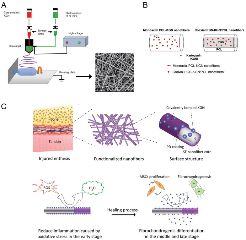
Figure 4.
(A) Schematic illustration of the fabrication process of KGN@PC nanofibrous scaffold. Reproduced with permission from (Yin et al., 2017) ©SAGE; (B) Schematic representation and monoaxial PCL-KGN fibers. Reproduced with permission from (Silva et al., 2020) ©elsivier; (C) Schematic diagram of integration and regeneration of bone-tendon interface by using a kartogenin- and polydopamine-functionalized silk fibroin nanofibrous scaffold. Reproduced with permission from (Chen et al., 2021). ©elsivier.

