. 2023 Sep 5;12:e81639. doi: 10.7554/eLife.81639

Figure 10. Comparison of RPA70N–peptide complex structures.

(A) Summary of the RPA70N-binding proteins for which RPA70N complex structures are available (Ddc2 binds to Rfa1N). (B) Superposition of apo RPA70N (orange, PDB: 2B29) with RPA70N structures determined in this study, colored using the peptide color code used in Figure 1F–N.

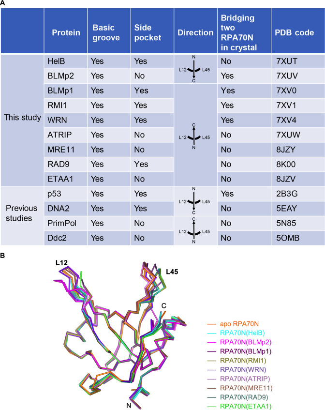
Figure 10—source data 1. Editable word file of Figure 10A.

Figure 10.

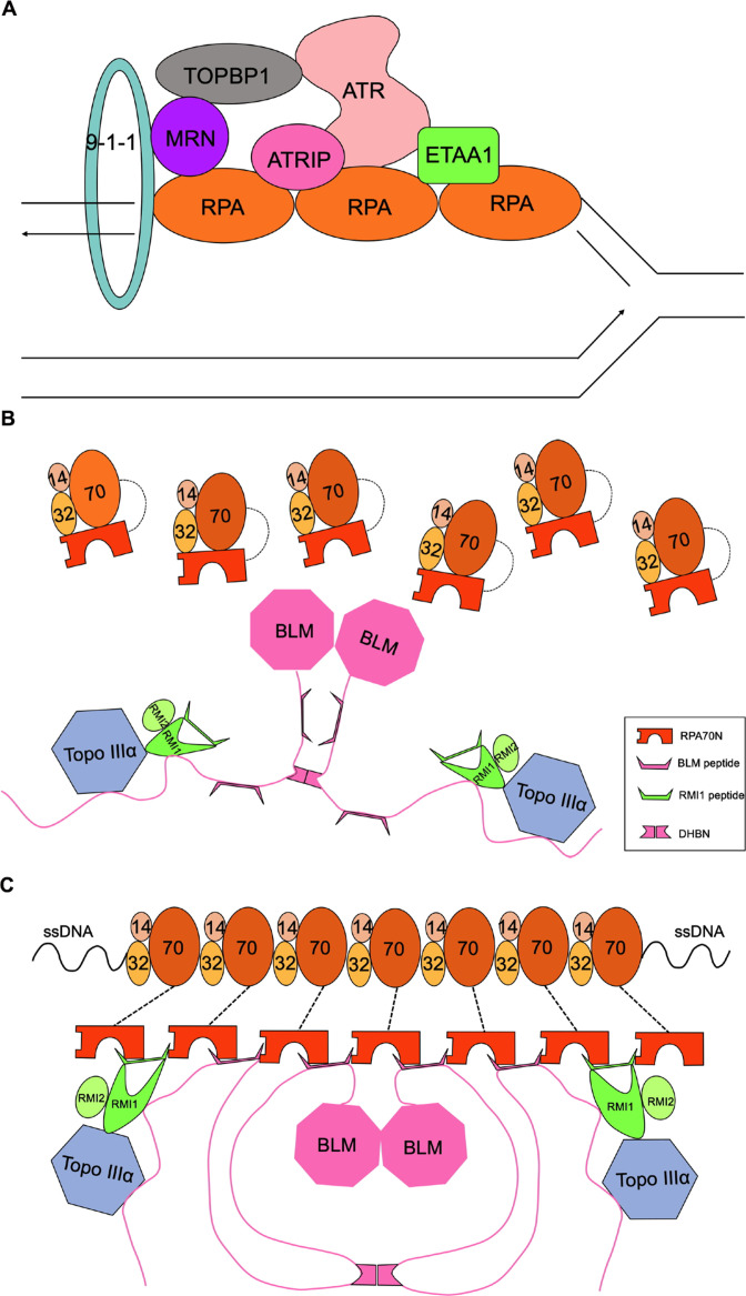
Figure 10—figure supplement 1. Model of protein recruitment in the RPA70N-mediated DNA damage response (related to Figure 10).

Figure 10—figure supplement 1.

(A) A simplified model showing how exposed ssDNA attracts RPA molecules, which recruit many DNA damage response proteins including ATR/ATRIP, the 9-1-1 clamp, MRN, ETAA1 and TOPBP1 to the stalled replication fork. (B) Without ssDNA, RPA molecules are not associated with the dimeric BTR complex because of the relatively weak affinity between RPA70N and the interacting sequences in the BTR complex. (C) Multiple RPA molecules nucleate on ssDNA, the increased avidity of RPA70N promotes BTR peptides to bind, and some bound peptides in turn promote RPA70N association by interacting with two RPA70N molecules simultaneously.