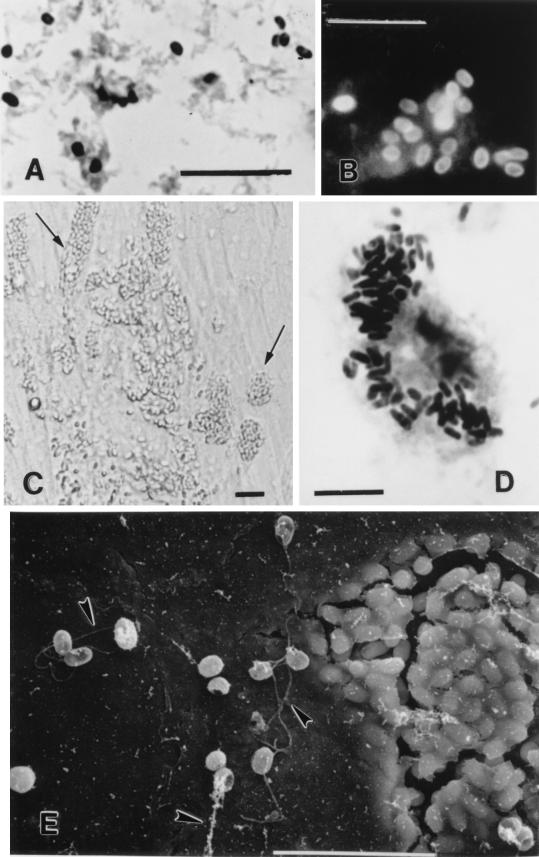
FIG. 1.
Optical and scanning electron microscopic images of microsporidia spores after treatment by various procedures. (A) Stool smear stained by the quick-hot Gram-chromotrope technique. Bar, 10 μm. (B) Stool smear from the same patient whose stool smear was used in panel A reacted with the anti-E. intestinalis serum. Bar, 10 μm. (C) Growth of E. intestinalis in cell culture. Note the host cells filled with spores (at the arrows). Differential interference contrast optics were used. Bar, 5 μm. (D) Smear of the culture supernatant from the same flask used for panel C but stained by the quick-hot Gram-chromotrope technique. Note the cell filled with darkly staining spores. Bar, 10 μm. (E) Scanning electron microscopic appearance of E. intestinalis from cell culture. Note the delicate thread-like polar tubules at the arrowheads. Bar, 10 μm.

