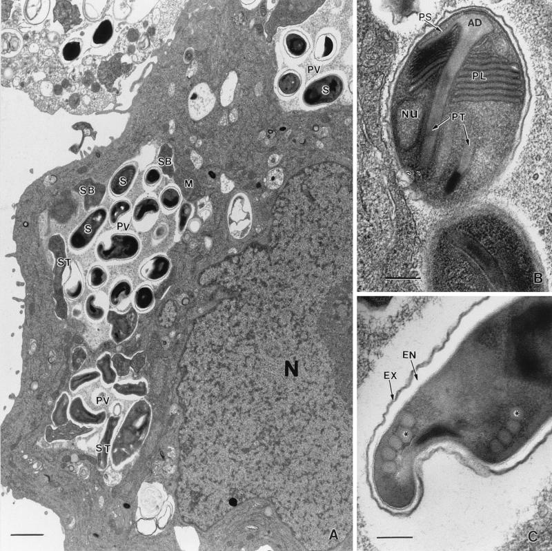
FIG. 2.
Ultrastructure of E. intestinalis within host cells. (A) Infected E6 cell demonstrating the classical septated parasitophorous vacuole (PV), a characteristic feature of E. intestinalis, filled with spores (S). M, meront; SB, sporoblast; ST, sporont; N, host cell nucleus. Bar, 1 μm. (B) Spore demonstrating a lamellar polaroplast (PL), a polar tubule (PT) with the anchoring disk (AD), and a nucleus (Nu). PS, polar sac. Bar, 200 nm. (C) Spore with four to five turns of the polar tubule (at the asterisk). EX, exospore; EN, endospore. Bar, 200 nm.

