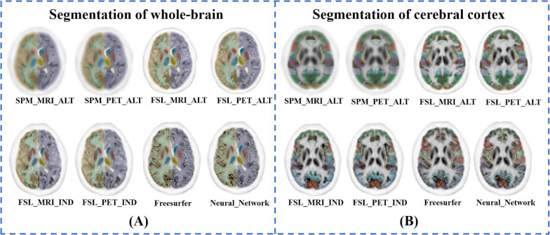
Fig. 3.
The segmentation results of different methods overlaid on the PET images of one subject. A The results at the whole-brain level, and B depicts the results at the cerebral cortex level. Different methods produced brain segmentations with similar appearances. The methods based on SPM and FSL divided the cortical brain regions into block-shaped areas that contain cerebrospinal fluid; in contrast, the method based on FreeSurfer and Neural Network divided the cortical brain regions into surface-shaped areas that do not contain cerebrospinal fluid

