Fig. 2.
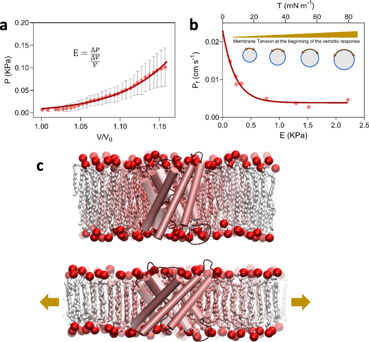
Experimental evidence demonstrates mechanosensitivity in AQPs. a Cell mechanics can be studied in Xenopus oocytes by the simultaneous measurement of pressure and volume during the osmotic response. The pressure-volume relationship evidences that the volumetric elastic module (E) rises as volume increases (Ozu et al., 2013). Dots represents the mean ± sem registered in BvTIP1;2-expressing oocytes during the osmotic response driven by a 100 mOsmol Kgw−1 gradient. Continuous line is illustrative, just to emphasize the nonlinearity of the response. Similar results were reported in Goldman et al. (2017). b By setting different membrane tension states at the beginning of the osmotic response (driven by the same osmotic gradient) demonstrates that the water permeability coefficient (Pf) decreases as the initial membrane tension increases. Dots represent independent experiments performed with BvTIP1;2-expressing oocytes. The osmotic gradient was the same in all the experiments. Figure modified from Goldman et al. (2017) under license permission from the publisher. The continuous line is illustrative, just to emphasize the non-linear decreasing relationship. c The lipid-protein interaction and the ordering of lipids around aquaporins were studied by both experimental and simulation approaches. Reported data suggest that there is not a fixed shell of lipids around AQPs. Instead, a dynamic interchanging occurs between this annular shell and bulky lipids (Stansfeld et al., 2013). This is represented by the lipid color gradient. The membrane tension increment (arrows in lower panel) produces thinning of the membrane. Therefore, hydrophobic regions of the protein tend to be exposed to the aqueous solution. Then, as occurs in MS ion channels, the hydrophobic mismatch is solved by a protein conformational change. Both the protein and the membrane are schematic representations with general purposes only

