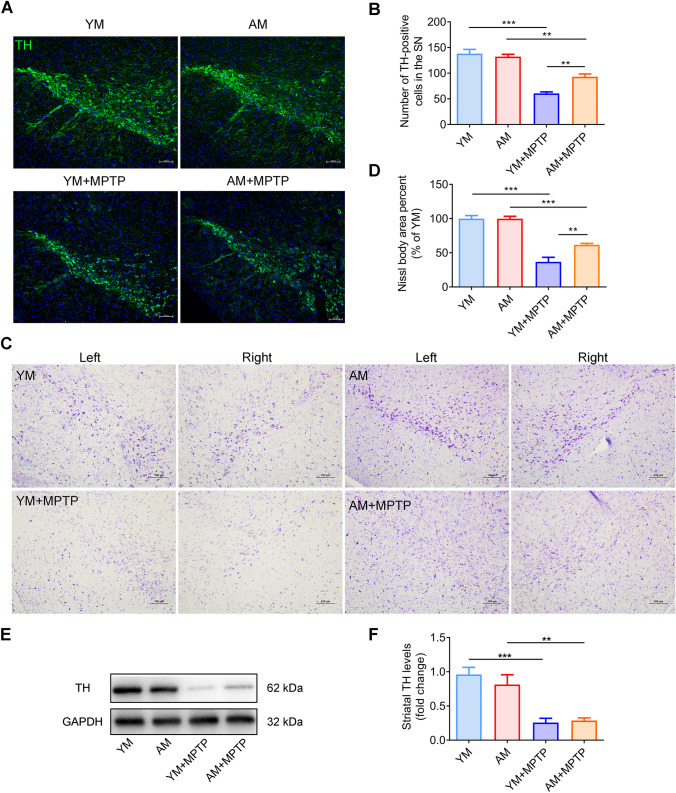
Fig. 4.
Effect of FMT from young and aged mice on brain dopaminergic neurons and TH expression. A Immunostaining of TH in the SNpc region of mouse brains in the different groups. B Quantification of TH-positive neuronal cell bodies with representative immunofluorescence images of SNpc sections. C Nissl staining of the SN of mice in different groups under the microscope. D Quantification of Nissl body area percent, as exhibited in (C). N = 5 in each group. Scale bar: 100 μm. E Western blotting for TH protein expression. F Quantification of TH expression in the striatum. N = 7 in each group. *: P < 0.05; **: P < 0.01 and ***: P < 0.001

