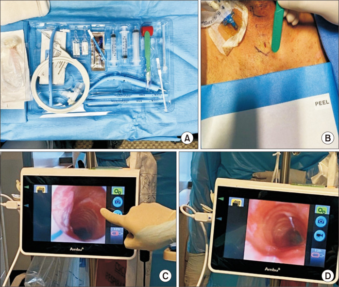
Fig. 1.
(A) The G2 Blue Rhino set from Cook Medical. (B) Once the patient was appropriately positioned and prepped, landmarks were identified and an 8-mm vertical incision was made over the cricoid cartilage. (C) Once the operator had dissected down to the trachea, the bronchoscope and endotracheal tube were withdrawn until the first three tracheal rings were visualized by confirming the triangulation of the subglottic trachea. (D) Once proper visualization was obtained, the introducer needle was placed, ideally anywhere between the first and third tracheal rings.

