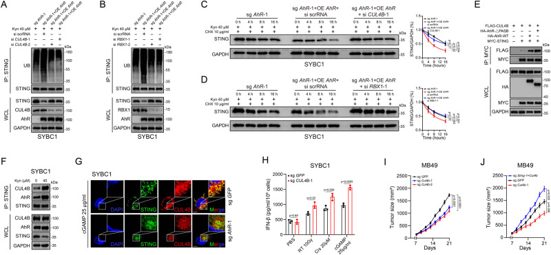
Fig. 6. AhR worked as an adaptor to facilitate STING ubiquitination through CUL4B-RBX1 E3 complex.
A, B Coimmunoprecipitation and immunoassay for extracts of cell lysate by anti-STING antibodies in CUL4B (A) and RBX1 (B) knocked-down cells. C, D Immunoassay for STING expression of AhR knocked-out cells with overexpressed AhR and knocked-down CUL4B (C) or RBX1 (D) after treated with Kyn in different time points. E Coimmunoprecipitation and immunoassay for extracts of HEK293T cells transfected with MYC-STING, HA-AhR (WT), AhR (1–277; 424–848), and FLAG-CUL4B by anti-MYC beads. F Coimmunoprecipitation and immunoassay for extracts of cell lysate after treated with Kyn by anti-STING antibodies. G Confocal microscopy for STING and CUL4B expression after being treated with cGAMP in AhR knocked-out cells. Scale bar 20 μm. H ELISA for IFN-β content in the supernatant of CUL4B knocked-out cells after indicated treatment. I Effect of Cul4b knocked-out on MB49 growth (n = 6) (Mean ± SEM). J Effect of Sting/Cul4b knocked-out on MB49 growth (n = 6) (Mean ± SEM). P value by one-way ANOVA (C, D, I, J). P value by two-tailed t test (H). All p value < 0.05 as statistic difference. Error bars represent Mean ± SD, unless otherwise indicated. Three biologically independent experiments were performed (A–H). Source data are provided as a Source Data file.

