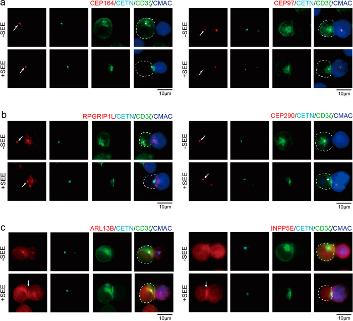
Fig. 1. Localization of cilium-associated proteins in resting and activated T cells.
Immunostaining of a proteins at the centriole distal end (CEP97, CEP164), b transition zone proteins (RPGRIP1L, CEP290), and c cilium-enriched proteins (ARL13B, INPP5E) in conjugates of Jurkat T cells (Left) and CMAC-labeled Raji B cells (Right), in the presence (+) or absence (–) of SEE. Cells were co-stained with anti-centrin 2 (CETN, cyan) and anti-CD3ζ (green) antibodies as centriole and immune synapse markers. Arrows indicate the localization of each cilium-associated protein. Images are representative of at least three independent experiments. T cells are depicted in dotted lines. Scale bar: 10 μm.

