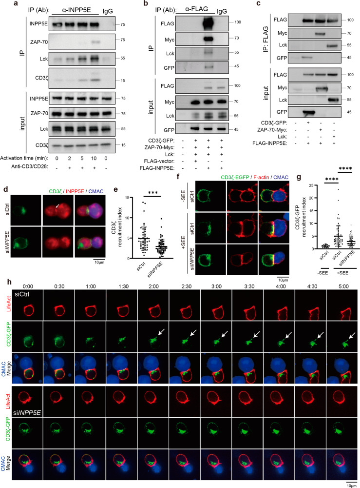
Fig. 4. INPP5E is required for CD3ζ recruitment to the immune synapse.
a Jurkat T cells were activated by crosslinking with the anti-CD3/CD28 antibodies for 2–10 min. The total cell lysates from each time point were immunoprecipitated (IP) with anti-INPP5E antibody. The immunoprecipitates were resolved by 12% SDS-PAGE followed by immunoblotting with indicated antibodies. Representative images are shown. IgG lane was used here as the control. b, c HEK293T cells were transfected with indicated plasmids. Cells were lysed and immunoprecipitated (IP) with the anti-Flag antibody. The immunoprecipitates were resolved by 10% SDS-PAGE followed by western blot with indicated antibodies. d Jurkat cells were transiently transfected with either siCtrl or siINPP5E. Immunostaining of CD3ζ and INPP5E in conjugates of Jurkat T cells and CMAC-labeled SEE-pulsed APCs. Scale bar: 10 μm. e Quantification of CD3ζ recruitment at immune synapse from (c). n = 46 conjugates for the siCtrl, and n = 49 conjugates for siINPP5E. Scale bar = 10 μm. Error bars indicate mean ± SD. Unpaired student T-test. ***P < 0.001. Arrows indicate localization of INPP5E. f Jurkat cells were transfected CD3ζ-GFP together with either siCtrl or siINPP5E. Immunostaining of CD3ζ-GFP in conjugates of Jurkat T cells and CMAC-labeled APCs, in the absence (–, upper) or presence (+, middle) of SEE. Cells were conjugated for 10 min, and co-stained with phalloidin to visualize F-actin. Upper and middle, control siRNA transfected cells; lower, siINPP5E transfected cells. Scale bar = 10 μm. g The recruitment index of CD3ζ-GFP at the immune synapse was quantified. Dotted black squares indicate the regions that were selected for quantifying the GFP fluorescence intensity. N = 4. n = 30 conjugates for siCtrl -SEE, n = 81 conjugates for siCtrl +SEE, and n = 73 conjugates for siINPP5E. Error bars indicate mean ± SD. One-way ANOVA analysis. ****P < 0.0001. h Jurkat cells were transfected with either siCtrl or siINPP5E at day 1, while CD3ζ-GFP and LifeAct-TagRFP are transfected at day 3. Cells were analyzed at day 4. Live images of cells conjugates were recorded for 5 min. Arrows indicate the localization of CD3ζ-GFP at the T-cell-APC contact site. The represented images are shown. The results were from 3–5 cells for each condition from two independent experiments.

